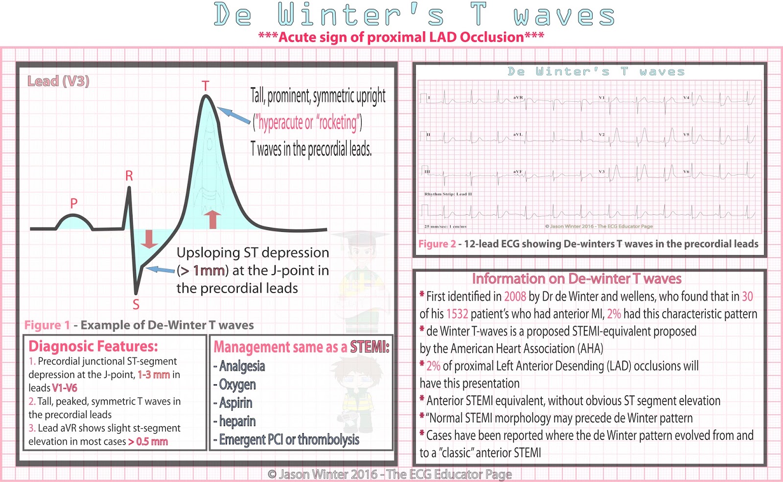
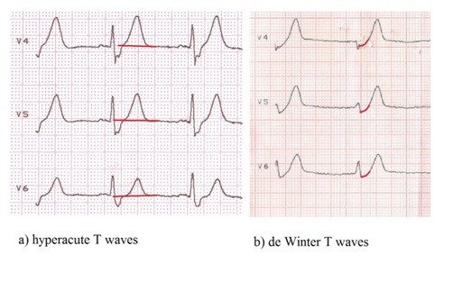
De Winter T Wave - File:De Winter's T wave (ECG).svg - Wikimedia Commons - Upsloping st depression and peaked t waves in precordial leads.. A new ecg sign of proximal lad occlusion. Tachycardia tall r wave in v1 tamponade tca tca overdose tca toxicity terminal qrs distortion thrombolytics torsades torsades de points toxicology tremor artifact trigeminy troponin twi. De winter t waves are a rare presentation for patients with acute lad occlusion, some authors considering this pattern to be an equivalent of anterior st elevation myocardial infarction (stemi)1. First reported by first reported de winter in key diagnostic features include st depression and peaked t waves in the precordial leads. While originally described as an early, static, and unique finding, the de winter t wave pattern has subsequently been demonstrated to precede classic st segment elevation, as well as occur in other coronary lesions, including left main coronary artery occlusion.
First identified in 2008 by dr. Cardiovascular implications of covid 19 by pranoti hiremath md. Descriptionde winter's t wave (ecg).svg. It is an atypical presentation of acute myocardial infarction due to lad occlusion. An ecg pattern not to miss‼ walksinto ed with sudden cp 1st #ecg with #stemi but then.
De Winter ST/T-Waves - ECG Medical Training from www.ecgmedicaltraining.com The aim of this case report was to highlight the dilemma in the management of a patient with de winter. De winter t waves are seen in 2% of left anterior descending coronary lesions. While originally described as an early, static, and unique finding, the de winter t wave pattern has subsequently been demonstrated to precede classic st segment elevation, as well as occur in other coronary lesions, including left main coronary artery occlusion. Descriptionde winter's t wave (ecg).svg. A new ecg sign of proximal lad occlusion. De winter sign on ecg is detected in 2% of patients with critical stenosis or occlusion of the left anterior descending artery. Changes in the structure of the heart and its surroundings (including blood composition) change the patterns of these four entities. The de winter ecg pattern is an anterior stemi equivalent that presents without obvious st segment elevation.
Depressions that indicate acute anterior.
An ecg pattern not to miss‼ walksinto ed with sudden cp 1st #ecg with #stemi but then. De winter sign on ecg is detected in 2% of patients with critical stenosis or occlusion of the left anterior descending artery. Cardiovascular implications of covid 19 by pranoti hiremath md. 2% of proximal lad occlusions will have this presentation. Tachycardia tall r wave in v1 tamponade tca tca overdose tca toxicity terminal qrs distortion thrombolytics torsades torsades de points toxicology tremor artifact trigeminy troponin twi. New england journal of medicine. The aim of this case report was to highlight the dilemma in the management of a patient with de winter. De winter's t waves were first described by de winter in an article to the editor of the new england journal of medicine in 2008(1). De winter t waves are seen in 2% of left anterior descending coronary lesions. De winter pattern on the ecg is associated with occlusion of proximal left anterior descending artery. The de winter pattern is seen in ~2% of acute lad. De winter t waves are a rare presentation for patients with acute lad occlusion, some authors considering this pattern to be an equivalent of anterior st elevation myocardial infarction (stemi)1. First reported by first reported de winter in key diagnostic features include st depression and peaked t waves in the precordial leads.
We present the case of a 41 years old male presenting with a 3 hours continuous chest pain. 2% of proximal lad occlusions will have this presentation. In 2008 described a new electrocardiographic sign of proximal lad occlusion, that consists of st segment upsloping depression at the j point in leads v1 to v6 that continued into tall, positive symmetrical t waves, that has been associated with the occlusion of the proximal left anterior. Descriptionde winter's t wave (ecg).svg. The de winter pattern is seen in ~2% of acute lad.
ECG Educator Blog : De Winters T waves from 1.bp.blogspot.com De winter sign on ecg is detected in 2% of patients with critical stenosis or occlusion of the left anterior descending artery. The aim of this case report was to highlight the dilemma in the management of a patient with de winter. 2% of proximal lad occlusions will have this presentation. We present the case of a 41 years old male presenting with a 3 hours continuous chest pain. These images are a random sampling from a bing search on the term de winter t wave. click on the image (or right click) to open the source website in a new browser window. The de winter ecg pattern is an anterior stemi equivalent that presents without obvious st segment elevation. While originally described as an early, static, and unique finding, the de winter t wave pattern has subsequently been demonstrated to precede classic st segment elevation, as well as occur in other coronary lesions, including left main coronary artery occlusion. Case study looking at the stemi equivalent of de winter t waves for lad occlusion.
Tachycardia tall r wave in v1 tamponade tca tca overdose tca toxicity terminal qrs distortion thrombolytics torsades torsades de points toxicology tremor artifact trigeminy troponin twi.
Tachycardia tall r wave in v1 tamponade tca tca overdose tca toxicity terminal qrs distortion thrombolytics torsades torsades de points toxicology tremor artifact trigeminy troponin twi. De winter's t waves were first described by de winter in an article to the editor of the new england journal of medicine in 2008 (1). New england journal of medicine. Written by salim rezaie 0 comments. The de winter ecg pattern is an anterior stemi equivalent that presents without obvious st segment elevation. The u wave represents papillary muscle repolarization. An ecg pattern not to miss‼ walksinto ed with sudden cp 1st #ecg with #stemi but then. De winter's t waves were first described by de winter in an article to the editor of the new england journal of medicine in 2008(1). In 2008 described a new electrocardiographic sign of proximal lad occlusion, that consists of st segment upsloping depression at the j point in leads v1 to v6 that continued into tall, positive symmetrical t waves, that has been associated with the occlusion of the proximal left anterior. De winter t waves are a rare presentation for patients with acute lad occlusion, some authors considering this pattern to be an equivalent of anterior st elevation myocardial infarction (stemi)1. De winter pattern on the ecg is associated with occlusion of proximal left anterior descending artery. The aim of this case report was to highlight the dilemma in the management of a patient with de winter. The de winter pattern is seen in ~2% of acute lad.
1) upsloping st elevation in avr (>= 1 mm) 2) st depressions and tall t waves in precordial leads #. New england journal of medicine. It is an atypical presentation of acute myocardial infarction due to lad occlusion. Depressions that indicate acute anterior. Represents an acute proximal occlusion (unlike wellen's sign which represents a subacute process).
ECG Rhythms: De Winter's ST/T ECG changes in huge anterior ... from 3.bp.blogspot.com Upsloping st depression and peaked t waves in precordial leads. The de winter ecg pattern is an anterior stemi equivalent that presents without obvious st segment elevation. Case study looking at the stemi equivalent of de winter t waves for lad occlusion. Represents an acute proximal occlusion (unlike wellen's sign which represents a subacute process). Descriptionde winter's t wave (ecg).svg. Original reports of the de winter pattern suggested that the ecg did not change or evolve until the culprit artery had been opened. First reported by first reported de winter in key diagnostic features include st depression and peaked t waves in the precordial leads. These images are a random sampling from a bing search on the term de winter t wave. click on the image (or right click) to open the source website in a new browser window.
The aim of this case report was to highlight the dilemma in the management of a patient with de winter.
We present the case of a 41 years old male presenting with a 3 hours continuous chest pain. Depressions that indicate acute anterior. While originally described as an early, static, and unique finding, the de winter t wave pattern has subsequently been demonstrated to precede classic st segment elevation, as well as occur in other coronary lesions, including left main coronary artery occlusion. The de winter ecg pattern is an anterior stemi equivalent that presents without obvious st segment elevation. Descriptionde winter's t wave (ecg).svg. De winter pattern on the ecg is associated with occlusion of proximal left anterior descending artery. Upsloping st depression and peaked t waves in precordial leads. De winter t waves are a rare presentation for patients with acute lad occlusion, some authors considering this pattern to be an equivalent of anterior st elevation myocardial infarction (stemi)1. The u wave represents papillary muscle repolarization. De winter's t waves were first described by de winter in an article to the editor of the new england journal of medicine in 2008 (1). The aim of this case report was to highlight the dilemma in the management of a patient with de winter. New england journal of medicine. Case study looking at the stemi equivalent of de winter t waves for lad occlusion.
Belum ada Komentar untuk "De Winter T Wave - File:De Winter's T wave (ECG).svg - Wikimedia Commons - Upsloping st depression and peaked t waves in precordial leads."

.svg - Wikimedia Commons - Upsloping st depression and peaked t waves in precordial leads.%20%2D%20https://miractsan.blogspot.com/2021/06/de-winter-t-wave-filede-winters-t-wave.html)
Belum ada Komentar untuk "De Winter T Wave - File:De Winter's T wave (ECG).svg - Wikimedia Commons - Upsloping st depression and peaked t waves in precordial leads."
Posting Komentar